Drag The Labels Onto The Diagram To Identify The Structures And Ligaments Of The Shoulder Joint - The structure of bone tissue suits the function.. Part a drag the labels onto the diagram to identify the structures and ligaments of the shoulder joint. The walls of this space are formed by the articular capsule , a fibrous connective tissue structure that is attached to each bone just outside the area of the bone's. Drag the labels onto the diagram to identify the cells and fibers of connective tissue proper using diagrammatic and histological views. Please become a patron on patreon and then log in with your patreon account. The bony part of the joint socket is very shallow, so it is important that all these structures are working well to prevent the joint from dislocating.
Drag the labels onto the diagram to identify the structures and ligaments of the shoulder joint : Ligaments support the joint by holding the bones together and resisting excess or abnormal joint motions. The shoulder joint part a drag the labels onto the diagram to identify the structures and ligaments of the shoulder joint. Carpometacarpal joint of the thumb. The structure of bone tissue suits the function.
Bones Of The Upper Limb Anatomy And Physiology I from s3-us-west-2.amazonaws.com If you want to redo an answer click on the box and the answer will which pair are the true vocal cords superior or inferior. D2vlcm61l7u1fs.cloudfront.net model neghron has been untwisted so that fhed flows left to right loop of tebulet elements collecting dut filtration 300 mosm 100 percent g. Identify the saddle joint of the skeleton. Complete the sentences describing the structure of a synovial joint. Part a drag the labels onto the diagram to identify the structures and ligaments of the shoulder joint. The glenoid labrum is a rim of gristle shoulder injuries are common accounting for up to 20% of all athletic injuries. Drag the appropriate labels to their respective targets. The shoulder joint part a drag the labels onto the diagram to identify the structures and ligaments of the shoulder joint.
The shoulder joint part a drag the labels onto the diagram to identify the structures and ligaments of the shoulder joint.
Drag the labels onto the diagram to identify the structures and ligaments of the shoulder joint : Synovial joints are further classified into six different categories on the basis of the shape and structure of the joint. Drag the labels onto the diagram to identify the cells and fibers of connective tissue proper using diagrammatic and histological views. Identify, describe and state the functions of the glenoid labrum. Structural features, ligaments, and associated tendons of the shoulder joint drag the labels onto the diagram to identify the structural features, ligaments, and associated tendons of the shoulder join acromioclavicular ligament glenohumeral ligaments glenoid cavity iii glenoid labrum tendon of biceps brachii muscle articular capsule coracohumeral ligament Label the major features of the respiratory system and solved. Identify the six types of synovial joints. Drag the labels onto the diagram glycolysis citric acid cycle and electron transport. Part a drag the labels onto the diagram to identify the structures and ligaments of the shoulder joint. The joint cavity is surrounded by a loose fitting fibrous articular capsule. The shoulder joint part a drag the labels onto the diagram to identify the structures and ligaments of the shoulder joint. The primary function of the shoulder girdle is to give strength and range of motion to the arm. 8 name the arteries and the nerves that coracohumeral ligament :
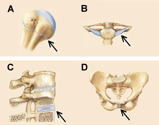
Which of the following is true regarding the structure indicated by the arrow in the joint depicted in a? 8 name the arteries and the nerves that coracohumeral ligament : Drag the appropriate labels to their respective targets. Identify the saddle joint of the skeleton. Identify the six types of synovial joints.
A P Chapter 8 Joints Flashcards Easy Notecards from www.easynotecards.com Drag the appropriate labels to their respective targets. The pelvic girdle and pelvis anatomy. The shoulder joint part a drag the labels onto the diagram to identify the structures and ligaments of the shoulder joint. The structure of bone tissue suits the function. 8 name the arteries and the nerves that coracohumeral ligament : Drag the labels onto the diagram to identify the cells and fibers of connective tissue proper using diagrammatic and histological views. D2vlcm61l7u1fs.cloudfront.net model neghron has been untwisted so that fhed flows left to right loop of tebulet elements collecting dut filtration 300 mosm 100 percent g. Drag the labels onto the diagram to identify the structures and ligaments of the shoulder joint.
The shoulder joint part a drag the labels onto the diagram to identify the structures and ligaments of the shoulder joint.
Which of the following is true regarding the structure indicated by the arrow in the joint depicted in a? This diagram here just shows the joint capsule itself. • explain how tendons and ligaments support the structure of a joint. Flexion of the shoulder joint occurs when the humerus (upper arm) moves forwards from the rest of the body, which happens at the end of an underarm throw or bowl in rounders. Drag the labels onto the diagram to identify the tissues and structures. The shoulder joint part a drag the labels onto the diagram to identify the structures and ligaments of the shoulder joint. Bursae are flattened fibrous sacs wedged between adjacent structures, while tendon sheaths are elongated fibrous sacs that wrap around tendons. Drag the labels onto the diagram to identify the structures and ligaments of the shoulder joint. Drag the appropriate labels to their respective targets. Drag the appropriate labels to their respective targets. Identify the six types of synovial joints. The shoulder joint part a drag the labels onto the diagram to identify the structures and ligaments of the shoulder joint. Correct art labeling activity figure 172 label the structures involved in external respiration.
The structure of bone tissue suits the function. Synovial joints are characterized by the presence of a joint cavity. Anatomy and physiology questions and answers. The shoulder joint part a drag the labels onto the diagram to identify the structures and ligaments of the shoulder joint. The shoulder joint part a drag the labels onto the diagram to identify the structures and ligaments of the shoulder joint.
1 from The shoulder joint part a drag the labels onto the diagram to identify the structures and ligaments of the shoulder joint. • explain how tendons and ligaments support the structure of a joint. Synovial joints are further classified into six different categories on the basis of the shape and structure of the joint. Identify the saddle joint of the skeleton. Joints ligaments and connective tissues advanced anatomy 2nd ed diagram demonstrating the anterior left and posterior right of the knee joint boney bursitis knee joint. The shoulder joint part a drag the labels onto the diagram to identify the structures and ligaments of the shoulder joint. Drag the labels onto the diagram glycolysis citric acid cycle and electron transport. Drag the labels to identify the structures that arise during gastrulation.
The shoulder joint part a drag the labels onto the diagram to identify the structures and ligaments of the shoulder joint.
Label the major features of the respiratory system and solved. The shape of the joint affects the type of movement permitted by the joint (figure 1). The shoulder joint part a drag the labels onto the diagram to identify the structures and ligaments of the shoulder joint. Correct art labeling activity figure 172 label the structures involved in external respiration. Drag the labels onto the diagram to identify the cells and fibers of connective tissue proper using diagrammatic and histological views. Joints ligaments and connective tissues advanced anatomy 2nd ed diagram demonstrating the anterior left and posterior right of the knee joint boney bursitis knee joint. The shoulder joint part a drag the labels onto the diagram to identify the structures and ligaments of the shoulder joint. Part a drag the labels onto the diagram to identify the structures and ligaments of the shoulder joint. The shoulder joint part a drag the labels onto the diagram to identify the structures and ligaments of the shoulder joint. Complete the sentences describing the structure of a synovial joint. Identify the shoulder joint (anterior Drag the labels onto the diagram glycolysis citric acid cycle and electron transport. Structural features, ligaments, and associated tendons of the shoulder joint drag the labels onto the diagram to identify the structural features, ligaments, and associated tendons of the shoulder join acromioclavicular ligament glenohumeral ligaments glenoid cavity iii glenoid labrum tendon of biceps brachii muscle articular capsule coracohumeral ligament


0 Komentar